Radiography in Akshayanagar, Bangalore
Orthopedic X-ray imaging support for accurate diagnosis, fracture assessment, joint evaluation, treatment planning, and follow-up care with Dr. Veeresh Patage.
Book AppointmentAbout Doctor
Dr. Veeresh Patage - Orthopedic Imaging and Diagnosis
- Radiography support for fracture diagnosis, joint pain, arthritis, deformity, sports injuries, and trauma assessment.
- X-ray review for bone alignment, healing progress, implant position, joint narrowing, and treatment planning.
- Integrated orthopedic consultation with imaging interpretation and next-step care guidance.
Dr. Veeresh Patage
MBBS, MS Orthopedics, Fellowship in Arthroplasty and Arthroscopy with 15+ years of orthopedic and trauma care experience.
Dr. Veeresh Patage uses radiography as a key part of orthopedic diagnosis, fracture evaluation, surgical planning, and follow-up care.
X-rays help identify bone breaks, joint degeneration, deformity, dislocation, implant position, and healing progress after treatment.
The focus is on accurate diagnosis, clear explanation, timely treatment planning, and monitoring recovery through appropriate imaging.

Dr. Veeresh Patage
Orthopedic surgeon using radiography and imaging review for fracture diagnosis, joint pain evaluation, arthritis care, trauma planning, and follow-up treatment.
Diagnostic Imaging
Advanced Radiography Services by Dr. Veeresh Patage
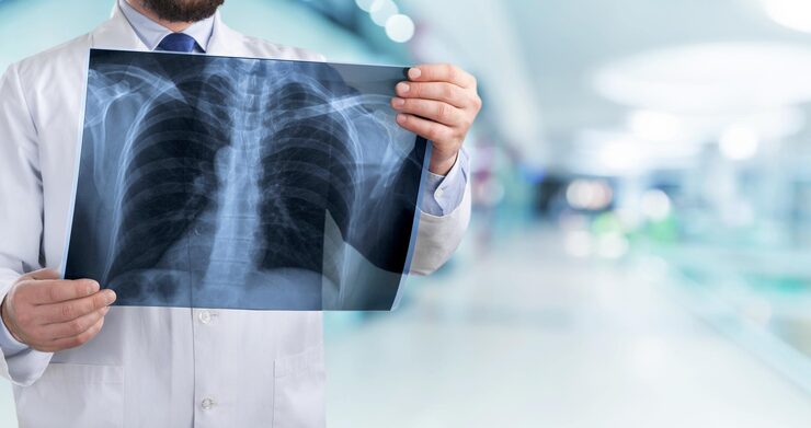
Radiography, commonly called X-ray imaging, is one of the most important tools in orthopedic diagnosis. It helps detect fractures, dislocations, arthritis, bone alignment problems, deformity, and healing progress.
Dr. Veeresh Patage provides radiography-based orthopedic evaluation in Akshayanagar, Bangalore to support accurate diagnosis, fracture care, joint treatment, surgical planning, and follow-up decisions.
Need X-ray or Report Review?
Share your X-rays, scans, symptoms, or injury details with the clinic team and plan evaluation with Dr. Veeresh Patage.
View Contact DetailsAccurate Imaging
X-ray views selected based on injury, pain location, and suspected diagnosis.
Orthopedic Review
Doctor-led interpretation for fractures, arthritis, alignment, and implant position.
Treatment Planning
Imaging findings connected with symptoms, examination, and care decisions.
Follow-up Care
Repeat imaging when needed to monitor healing, alignment, and recovery.
Understanding Radiography
What is Radiography?
Radiography is an imaging technique that uses X-rays to create pictures of bones and joints. It is commonly used in orthopedics to diagnose fractures, arthritis, dislocations, deformities, and healing progress.
X-ray findings are interpreted along with symptoms and clinical examination to decide the right treatment plan.
Diagnostic Uses
When Radiography is Used
Fracture Diagnosis
Detecting broken bones, alignment, and joint involvement.
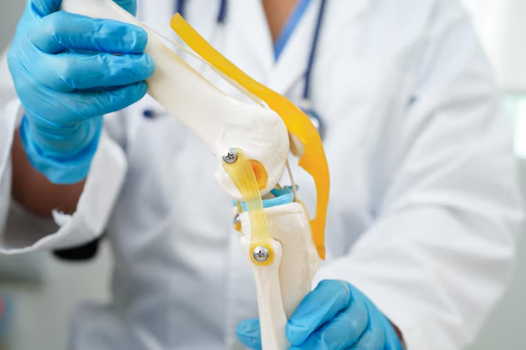
Joint Evaluation
Assessing arthritis, joint narrowing, deformity, and degeneration.
Healing Follow-up
Monitoring bone healing, implant position, and alignment.
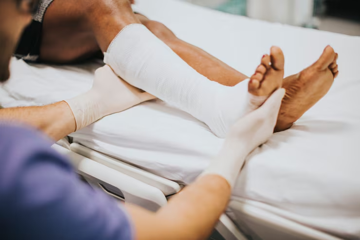
Injury Assessment
Evaluating pain after falls, sports injuries, and accidents.
When to Consult
When is Radiography Required?
X-rays may be recommended after trauma, fall, swelling, deformity, persistent pain, suspected fracture, arthritis symptoms, joint stiffness, or postoperative follow-up.
- Pain, swelling, deformity, or inability to bear weight after injury.
- Persistent joint pain, stiffness, arthritis symptoms, or reduced movement.
- Follow-up after fracture treatment, surgery, or implant placement.
Recovery Path
Imaging and Review
The required X-ray views are selected based on the affected body part and suspected condition. The images are then reviewed with the patient's symptoms and examination findings.
If X-rays are not enough for complex injuries, further imaging may be advised depending on the diagnosis.
Patient Outcomes
Benefits of Radiography
- Quick diagnosis of fractures, dislocations, arthritis, and bone alignment issues.
- Better treatment planning for casts, surgery, rehabilitation, or follow-up care.
- Clear monitoring of bone healing and implant position over time.
Common Questions
Frequently Asked Questions

Our Services

Advanced Orthopedic Care
For Better Mobility
Robotic Knee Replacement
Advanced Mako robotic technology for precise knee replacement with faster recovery and better outcomes.
Robotic Hip Replacement
Minimally invasive robotic-assisted hip surgery ensuring accuracy, reduced pain, and improved mobility.
Joint Replacement
Comprehensive solutions for damaged joints including knee, hip, and shoulder replacements.
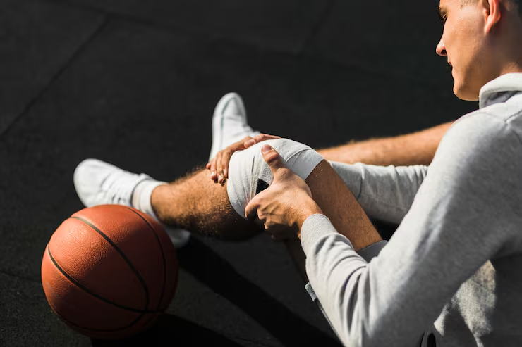
Sports Injury Treatment
Expert care for ligament tears, sprains, and sports-related injuries with faster return to activity.
Knee Replacement
Effective treatment for severe knee arthritis to restore movement and reduce pain.
Hip Replacement
Advanced surgical solutions for hip joint damage, improving mobility and quality of life.
Fracture Management
Comprehensive care for simple and complex fractures with modern surgical techniques.
Pediatric Fracture Treatment
Specialized care for children's fractures ensuring proper healing and growth.
Consult Dr. Veeresh Patage for Radiography in Akshayanagar
-
Call Now
+91 7019171578 -
Location
Dr. Veeresh Patage, Akshayanagar